
X-Ray (Radiography) is a form of light or electromagnetic radiation that is mainly used for medical imaging. This post will discuss in detail about what is X-Ray Radiography, types of imaging using X-Ray, how it works, applications, advantages and Disadvantages.
What is X-Ray (Radiography)
Like Visible light, X-Rays are also part of the electromagnetic spectrum. However, unlike visible light, they have higher energy and shorter wavelength. It was actually an accidental discovery by a German Mechanical Engineer Wilhelm Conrad Roentgen in 1895.

Fig. 1 – Introduction to X-RayÂ
It is also called the Roentgen Radiation in honor of the German Engineer, who went on to win the 1901 Nobel Prize in Physics. Nikola Tesla, the great inventor, is known to have been working with X-Rays in 1894 and had shared some of his images with Roentgen in 1895, who himself was impressed by Tesla’s work.
The X-Ray machine in hospitals captures body images that assist medical diagnosis and intervention. The wavelength of X-Ray ranges from 10 picometres to 10 nanometres.
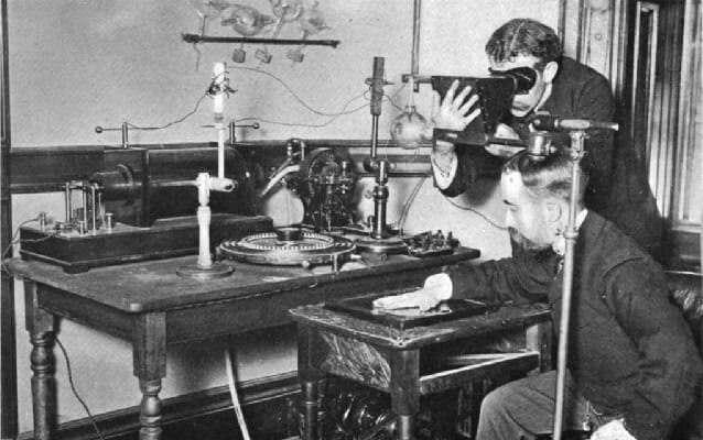
Fig. 2 – William Roentgen and the First X-Ray (Radiography)
Types of Imaging using X-Ray
The different types of Imaging using X-Rays are listed below:
- Portable X-Ray Machine
- Mammography Machine
- CT Scan or Computed Tomography Scan
- C-arm Machine
Portable X-Ray Machine
The Portable X-Ray Machine is equipped with a Tungsten filament which produces electrons when heated. These electrons move speedily down the tube and hit a metal electrode to produce X-Rays. This machine is light, user-friendly, needs less processing time, reduces radiation exposure and its high-tech features ensure high-quality images. Its cost-effectiveness and versatility ensure that it remains in great demand in the healthcare industry. This machine is particularly helpful for those who are critically ill or whose movements are restricted. It is in popular demand in nursing homes, home health care, forensic labs and dental camps.
Mammography Machine
The Mammography Machine (also called Mastography) is used to detect and diagnose early stages of breast diseases. Inside the machine is a tube which produces x-rays. This machine is designed to take images of breast tissues only; low energy x-rays do not generate clear pictures, hence a device is used to flatten the breast so that images can be captured from different angles. Today we have the benefits of:
- Digital Mammography: Clear images with low doses of x-rays.
- Computer-aided Detection: Highlights the abnormal areas.
- Breast Tomosynthesis: Cancer detection rates have improved.
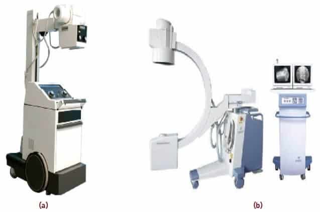
Fig. 3 – Types of X-Ray Machines (a) Portable Radiography Machine (b) C-arm Radiography Machine
CT Scan or Computed Tomography Scan
CT scan or Computed Tomography scan produces cross-sectional images of bones, blood vessels and soft tissues with the help of computer processing. In the conventional X-Ray machine, a fixed tube emits rays in only one direction. But in the CT scan narrow beams of X-Rays rotate around the patient; they are picked up by the detectors and transmitted to the computer. These images show more detail than a conventional X-Ray. The doctor can plan procedures for various treatments or in case of Tumours the progress achieved after radiation treatment.
C-arm Machine
C-arm is a complex X-Ray machine which is used whenever a patient needs to be imaged from multiple angles. In this scenario, the patient usually lies down and the C-arm moves around him to capture the images.
How does X-Ray Machine Work
An X-Ray machine is made up of tube-like structure that consists of a cathode and an anode. The cathode, a filament, releases energy in the form of electrons when introduced with an electric current. These electrons are attracted to the Anode, a Tungsten disc, on the opposite end of the tube. When the electrons come in contact with Tungsten, energy is released in the form of Photons. This energy then passes through a series of filters in a lead cylinder creating an X-Ray beam.
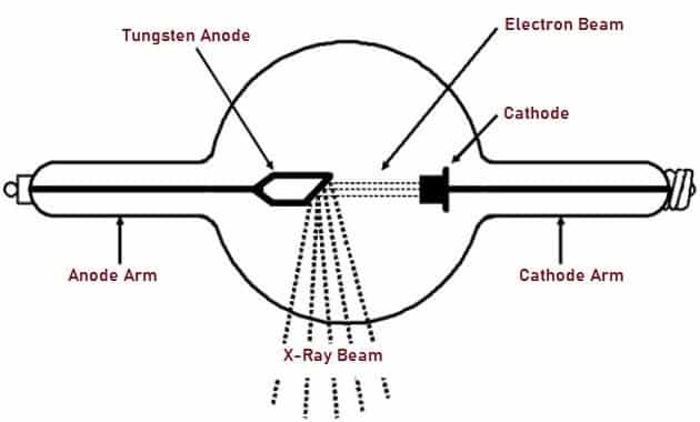
Fig. 4 – Schematic Representation of Typical X-Ray Tube
This beam is directed towards the body part which needs to be imaged. The patient is kept between the machine and the film/X-Ray detector. The dense body tissues, like bones, absorb this beam. When the Rays come in contact with the film, a chemical reaction takes place revealing the image.
Applications of X-Ray
Applications include:
- Radiography is an important diagnostic tool hence its application covers a wide network of medical science. The following are the four branches in which Radiology plays a major role in the diagnosis and treatment of the patient.
- Orthopaedic Radiology or Musculoskeletal Radiology helps in detecting the exact location of the injury, to bones, joints, muscles, ligaments and cartilage. It helps the doctor to assess the injury and plan a proper line of treatment.
- Dental X-rays help to diagnose the oral health of a person who has symptoms of oral disease. Different types of X-Ray imaging present different views of the mouth. They are:
- Radiation therapy is the use of X-Rays to kill Cancer cells or shrink the Tumour. Low energy X-Rays are used for treating skin cancer while high energy beams are used to treat cancer inside the body. Radiation therapy improves the survival rate of patients.
- Fluoroscopy is like a “movie†because real-time moving images within the body are captured by the X-Ray beam which transmits these moving images to a screen. A contrast dye highlights the affected part over which the X-Ray beam passes and images are captured. Fluoroscopy is used in procedures like Barium X-Rays and Arthrography.
Advantages of X-Ray
Advantages are:
- X-Ray Radiography can easily detect bone fractures, infections, calcifications and tumours.
- Quick diagnosis is possible because the X-Ray machine generates images very quickly.
- It can easily locate foreign objects in the body, thus paving the way for targeted medical intervention.
- The portable X-Ray machine can be wheeled into operation theatres, hospital wards, ICU’s and nursing homes.
- As it helps in the easy detection of tumours, invasive surgery can be avoided.
Disadvantages of X-Ray
Disadvantages include:
- X-Rays are considered carcinogenic by the WHO standards.
- Early technicians had suffered from burns and dry and flaky skins in their hands as they were not shielded from the Radiation.
- Large doses of Radiation used in CT and Fluoroscopy procedures raise the risk of cancer to the patient.
- The foetus runs a risk of Radiation hazard if a pregnant woman undergoes X-Ray investigation.
- The risk of cancer is high when X-Rays increase Hydrogen Peroxide levels in our blood.
- Patients exposed to high Radiation (Chemotherapy) suffer from multiple side effects like vomiting, loss of hair, fainting spells and bleeding.
The Future
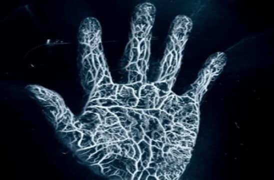
Fig. 5 – Image of a Hand Captured Using Photoacoustic Imaging
A new technique called Photoacoustic Imaging is being developed which uses laser-induced sound waves to capture images. This technology captures clear images of structures within our body with no discomfort or ionizing radiation involved.
Also Read:
Machine Learning - How it Works, Types, Applications, Advantages
Thomson’s Atomic Model – How it Works, Postulates and Limitations
Solar Power Satellite – How it works, Architecture, Application, Advantage
Its nice that you mentioned how portable x-ray machines are in great demand due to their cost effectiveness and versatility. I went to a nursing home the other day and saw a machine on the side, I discovered that it was actually a portable x-ray machine after I asked one of the nurses. It seems to be quite a convenient device to have and it wouldn’t hurt for any medical related place to have one.
Great read!!! Thanks for sharing such a great blog, blog like these are really helpful.
clm9ip
Embark into the vast galaxy of EVE Online. Find your fleet today. Create alongside hundreds of thousands of explorers worldwide. [url=https://www.eveonline.com/signup?invc=46758c20-63e3-4816-aa0e-f91cff26ade4]Start playing for free[/url]
Thanks for sharing. I read many of your blog posts, cool, your blog is very good.
A great overview of how widely X-rays are used across different industries.
Loving the info on this web site, you have done great job on the blog posts.
websites companies [url=https://otvetnow.ru]https://otvetnow.ru[/url] ftp site
Very nice article and right to the point. I am not sure if this is actually the best place to ask but do you guys have any ideea where to employ some professional writers? Thanks 🙂
It’s actually a nice and helpful piece of information. I am happy that you simply shared this useful information with us. Please stay us up to date like this. Thank you for sharing.
Thanks for sharing. I read many of your blog posts, cool, your blog is very good.
I like what you guys are up too. Such clever work and reporting! Keep up the excellent works guys I’ve incorporated you guys to my blogroll. I think it will improve the value of my site 🙂
I gotta favorite this website it seems extremely helpful extremely helpful
Wow! This can be one particular of the most helpful blogs We have ever arrive across on this subject. Basically Fantastic. I’m also a specialist in this topic therefore I can understand your effort.
wixt9o
j7n1vi
b35c9p
Excellent blog! Do you have any tips for aspiring writers? I’m planning to start my own website soon but I’m a little lost on everything. Would you recommend starting with a free platform like WordPress or go for a paid option? There are so many choices out there that I’m completely confused .. Any suggestions? Bless you!
I really appreciate this post. I have been looking everywhere for this! Thank goodness I found it on Bing. You have made my day! Thank you again
I am glad to be one of the visitors on this outstanding site (:, thanks for putting up.
I love your writing style truly loving this website .
Thank you for your sharing. I am worried that I lack creative ideas. It is your article that makes me full of hope. Thank you. But, I have a question, can you help me?